What Is An Mri Of The Spine
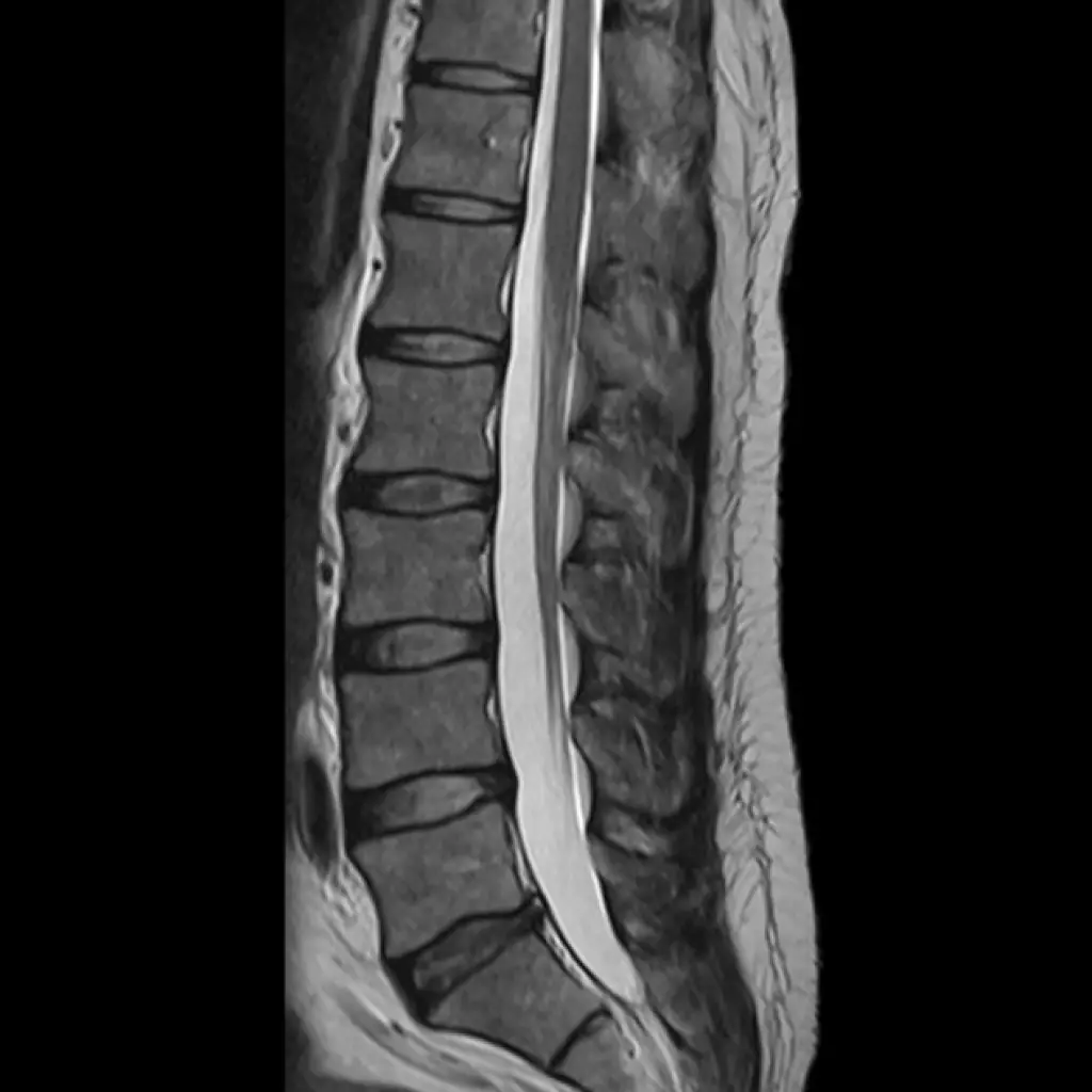
Magnetic resonance imaging uses a magnetic field and radio waves to take detailed images of your spine and nearby tissues. A spine MRI is useful in evaluating the soft tissue structures of the back, including the position of the vertebrae that make up your spinal column.
An MRI scan of the spine may spot abnormalities indicative of infection, nerve and disc problems, arthritis, blood vessel problems, and spinal tumors.
What Can A Thoracic Spine Mri Tell You
The thoracic segment includes 12 vertebrae, which are larger than the cervical vertebrae, and are part of the structures of your upper or middle back.
These structures include muscles, thoracic ligaments, tendons, and intervertebral discs. Your thoracic spine also includes 12 sets of ribs and the structures that make the thoracic cavity. The joints are attached with another type of soft tissue called cartilage.
Some abnormalities that may be spotted on a thoracic spine MRI are:
- Tumors in the spinal canal
- Tumors of the spinal cord
- Bulging spinal discs
- Abscesses and other signs of infection
Why Are Lumbar Spine Mris Done
A lumbar spine MRI can detect a variety of conditions in the lower back, including problems with the bones , soft tissues , nerves, and disks.
Sometimes, doctors order an MRI to check the anatomy of the lumbar spine or to look for injuries in the area. For example, the scan can find areas of the spine where the spinal canal is too narrow and might require surgery. It can assess the disks to see if theyre bulging, ruptured, or pressing on the spinal cord or nerves.
The MRI also can help doctors:
- Evaluate symptoms such as lower back pain, leg pain, numbness, tingling or weakness.
Recommended Reading: Where Do I Go For Lower Back Pain
What Is A Spinal Mri
A spinal MRI, or magnetic resonance imaging, uses powerful magnets, radio waves, and a computer to make clear, detailed pictures of your spine.
You may need this scan to check for spine problems, including:
-
Numbness, tingling, and weakness in your arms and legs
The MRI may scan your whole spine or just a part of it. Unlike X-rays and computerized tomography scans, it doesnât use damaging radiation. Itâs generally safe and painless. The doctor will let you know about any possible risks you may face. Theyâll also tell you if you canât get the procedure because of certain implants that you have.
Determining The Source Of Back Pain With Mri

In most cases, patients who have back pain have injured the muscles, ligaments or tendons that support the spinal column. If the pain is focused in the lower back, you may have an issue with the lumbar spine. Pain in the upper back or neck can often be attributed to a cervical spine condition. Back injuries are typically caused by poor posture, degenerative conditions, a physically demanding occupation, genetics, medical history, poor physical health, lack of exercise, or some combination of these factors.
Depending on the exact location and severity of your back pain, your doctor may initially recommend over-the-counter medications, physical therapy, or changing how you sit, move, or lift things before referring you for a diagnostic imaging study.
If conventional methods and lifestyle changes are not enough to alleviate symptoms or help treat the source of your pain, an MRI scan can be used to detect the source of back pain.
MRI or magnetic resonance imaging is an advanced technology that uses a magnetic field and radio waves instead of X-rays to produce high-quality images. MRI scans for back pain capture images of soft tissue, including the disks that separate the vertebrae, the spinal cord, the muscles and connective tissue around the spine, and the nerves that run in and out of the spine.
Recommended Reading: Can Diclofenac Gel Be Used For Back Pain
Should I Be Concerned About X
Everyone is exposed to naturally-occurring radiation and the amount varies depending on where you live. When you undergo an x-ray, the radiation that is not absorbed by your body creates your spinal image. Your radiation dose is the amount of radiation your body absorbs every time you undergo x-ray. Radiation dose to your entire body is measured as the millisievert also called the effective dose.
The value of effective dose helps your doctor measure your risk for potential side effects of undergoing radiographic imaging . For example, it is known certain body tissues and organs in the lower back regions are sensitive to radiation exposure such as the reproductive organs . Also, radiation exposure can increase the risk for cancer.³
The Anatomy And Function Of The Spine
Also called the backbone, the spinal column is a complex group of bones that creates your bodys main support. The bones of the spine are called vertebrae. You have 33 of them stacked on top of each other, interlocking to form the column that houses your spinal cord.
Between each vertebra are tiny shock absorbers called intervertebral discs. Ligaments are soft tissues that hold the vertebrae together, and tendons connect them to muscles. Your spine works with your nervous and musculoskeletal systems to make sure you can sit, twist, bend, and walk. Fun fact: A healthy spine actually has a natural S-shaped curve.
The five sections or segments of your spine are: cervical, thoracic, lumbar, sacral, and coccygeal. Lets look at why you might get an MRI of each part.
Recommended Reading: Can Ibuprofen Help Back Pain
What Can A Cervical Spine Mri Show
Your cervical spine, or C-spine, consists of the first seven bones or vertebrae of your spinal column.
A cervical spine MRI scans the neck region from the base of your head to the beginning of the thoracic or mid-back region. It includes structures like the thyroid gland, throat, larynx, neck muscles, ligaments, tendons, and other soft tissues.
A few types of abnormalities that a cervical spine MRI procedure may help detect include:
- Congenital birth defects
- Irregularities in the position of vertebrae or in the curvature of the spine
- Pinched nerves
- Cancer tumors of the cervical spine
- Thyroid gland tumors
- Spinal stenosis, which is a narrowing of the spinal column
- Aneurysm or weakened vessel wall
- Vascular malformations or abnormally developed blood vessels
- Bleeding
What To Expect With An Mri
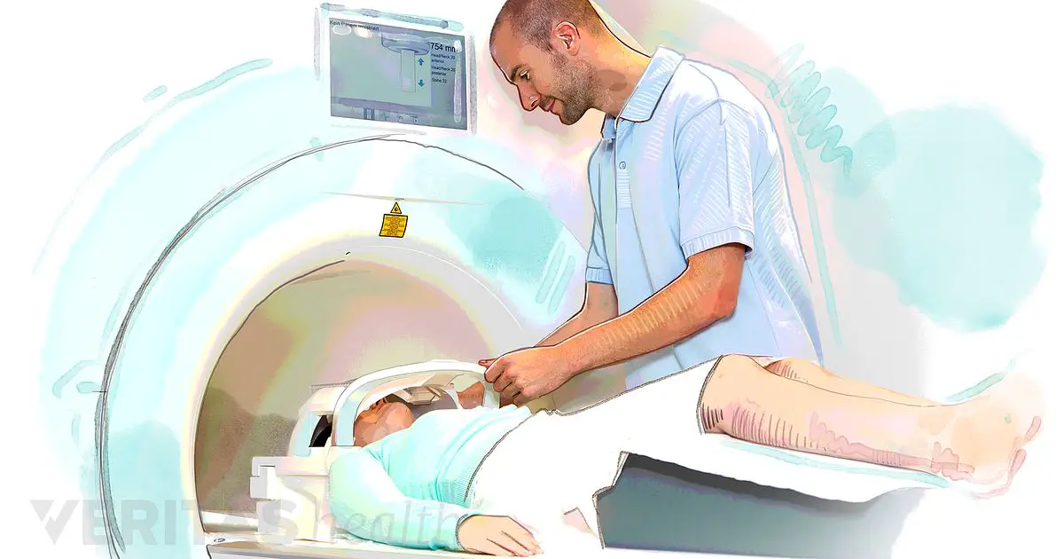
Well, basically you lie down on a table, and they slide you into a long tube with magnets humming in a creepy, err, consistent rhythm all around you. Its kind of like being in an episode of Twilight Zone. I have one word of warning, if you are claustrophobic, you may want to talk to your doctor and see if they can prescribe some kind of a relaxant to help. I will get more into that a little bit later.
Some people fall asleep during an MRI, they think the creepy noise of the machine is actually soothing and comforting I am not one of those people.
Sometimes the doctor will order the MRI with contrast dye to improve the quality of the images. An MRI may or may not be administered with contrast dye it just depends on what the doctor orders.
If you do need contrast, this is not a big deal, BUT the MRI tech will poke you with a needle so they can inject the dye during the exam. So if you have trypanophobia , you will want to prepare for that.
You also have to remain pretty darn still throughout the exam so that they can have clear images to look at. Imagine you are taking pictures, you need a steady hand, so the pictures are not blurry right?
Also Check: How To Use An Inversion Table For Lower Back Pain
What Matters Most To You
Your personal feelings are just as important as the medical facts. Think about what matters most to you in this decision, and show how you feel about the following statements.
Reasons to have an MRI
Reasons not to have an MRI
I’ve had low back pain for several months, and I want to find out if something serious is wrong.
I’m willing to give my low back pain more time to go away on its own, even if it takes a year.
Contact Ahi To Schedule Your Lumbar Mri
If your doctor orders an MRI for your back, contact AHI today. We offer the same high-quality scans using the newest and most up-to-date equipment and leading-edge processes at a cost thats usually significantly lower than hospitals or clinics. With 30 centers located in four states, AHI is both convenient and affordable.
Recommended Reading: How To Treat Back Pain
Should You Get An Mri For Low Back Pain
Have you considered getting an MRI as the missing piece of the puzzle in finally living a life without back pain?
Unfortunately, this is the sentiment we hear far too often from our clients living with chronic low back pain.The longer the pain continues, the more strongly you might consider an MRI as the best option.
How An Mri Works During A Scan

The MRI machine uses a strong magnetic field that aligns and stimulates particles called protons in the body, forcing them to spin out of alignment.
When the technician halts the magnetic field, the protons begin to spin in their usual way. As they do this, they give off energy that the MRI machine detects. The MRI machine records this information, and a computer processes the data to create a detailed image of the body area.
Also Check: Is Icy Hot Good For Back Pain
Low Back Pain: Mri Vs Ct Scan
The best test to obtain when looking at the spine is an MRI rather than the CT scan, says Dr. Michael Perry, MD, member of the North American Spine Society and American College of Sports Medicine.
These scans are the best for soft tissues, such as your spinal nerves, disc, cord and ligaments, continues Dr. Perry. An MRI will allow your doctor to see cord and nerve compression.
Thats not the greatest news to those who fear enclosed spaces and dont care about radiation.
But its really good news because you should be concerned about radiation, and the MRI is truly superior at imaging the causes of low back pain.
So if youre scared of being inside a tube, remind yourself that the MRI equipment does not emit radiation, and the CT scanner does.
When you may have a bone issue, such as a hairline fracture, spurring or arthritis, a CT scan is the best test to obtain, says Dr. Perry.
A CT scan is preferable if you have a pacemaker, defibrillator or morphine pump. When you have one of these implanted devices, an MRI scan is contraindicated because of the MRIs ability to interfere with the functions of these devices.
If youre scheduled for an MRI due so low back pain, bring good earplugs with you, as this machine produces loud knocking noises throughout the procedure.
Dr. Perry is chief medical director and co-founder of USA Spine Care & Orthopedics, and is frequently sought out for his minimally invasive spine surgery expertise.
What Can You Learn From A Lumbar Spine Mri
The lumbar spine is your lower back, but an MRI of the lumbar spine usually includes the sacral and coccygeal areas as well. Your lumbar spine has five vertebrae, and they are larger than those in the thoracic region. The thorax connects to your pelvis and sacrum. It includes large muscles that help you bend and lift and carry heavy loads. It also has a complex network of nerves and blood vessels.
During a lumbar MRI, some abnormalities that may show up are:
- Bulging or herniated disc
You May Like: How To Self Treat Lower Back Pain
Indications And Contraindications For An Mri Scan
The following general rules are usually considered by a physician before ordering an MRI scan for a patient with back pain, neck pain or leg pain stemming from a spine problem.
Indications for when to get an MRI scan include:
- After 4 to 6 weeks of leg pain, if the pain is severe enough to warrant surgery
- After 3 to 6 months of low back pain, if the pain is severe enough to warrant surgery
- If the back pain is accompanied by constitutional symptoms that may indicate that the pain is due to a tumor or an infection
- For patients who may have lumbar spinal stenosis and are considering an epidural injection to alleviate painful symptoms
- For patients who have not done well after having back surgery, specifically if their pain symptoms do not get better after 4 to 6 weeks.
Contraindications for undergoing an MRI scan for spine-related pain in the back, neck or leg include:
- Patients who have a heart pacemaker may not have an MRI scan
- Patients who have a metallic foreign body in their eye, or who have an aneurysm clip in their brain, cannot have an MRI scan since the magnetic field may dislodge the metal
- Patients with severe claustrophobia may not be able to tolerate an MRI scan, although more open scanners are now available, and medical sedation is available to make the test easier to tolerate
- Patients who have had metallic devices placed in their back can have an MRI scan, but the resolution of the scan is often severely hampered by the metal device and the spine is not well imaged.
When Should I Get An Mri Of My Low Back
MRI plays an especially vital role in helping your doctor know how to get rid of your low back pain. However, studies show getting an MRI too soon can lead to a bad result. So, when should you get an MRI of your low back?
Most people with low back pain have arthritis, compression fracture, a torn disc, herniated disc, or spinal stenosis . Arthritis and compression fractures show up on x-ray, but it takes MRI to know if you have a torn disc, herniated disc, or spinal stenosis. Your doctor will need to know which problem is present to recommend the right treatment plan. If you have red flags suggesting a serious underlying problem, you need an MRI and doctor visit emergently, which means right away.
But for most people, MRI should wait. The right time to get an MRI of your low back depends on how long your back has been hurting and the type of pain you experience.
To determine if you need an MRI of your back, you must know which of the five types of low back pain you have. Read to the end to learn to identify your pain and whether you need an MRI.
Your low back comprises five cube-shaped vertebral body bones separated by rubbery discs. Behind the bones, your spine forms a canal to contain and protect your nerve roots. Two facet joints connect each bone, one on each side, to the bones above and below except the fifthlumbar bone, which connects to your tailbone by two facet joints.
The facet joints can become inflamed and hurt just like any other joint in your body.
Also Check: Do Epidural Shots Work For Back Pain
Do I Need An Mri Scan
The MRI was developed in the 1980s and has revolutionized treatment for patients with low back pain. An MRI scan is generally considered to be the single best imaging study of the spine to help plan treatment for back pain.
Physicians usually have a good idea of what they are looking for on the MRI scan before one is performed. The scans are most commonly used for pre-surgical planning, such as for a decompression or a lumbar spinal fusion. MRI scans are extremely sensitive to picking up information about the health of the discs, as well as the presence of any spinal tumors or a lumbar disc herniation that pinches the nerve roots and causes back pain.
In addition to pre-surgical planning, MRI scans are also very useful for the following:
- To rule out infection or tumor
- For patients who have had back surgery, to differentiate scar tissue from a recurrent disc herniation.
- Prior to performing an epidural injection to rule out the risk of injecting a steroid into a tumor or infection
How A Lumbar Mri Is Performed
An MRI machine looks like a large metal-and-plastic doughnut with a bench that slowly glides you into the center of the opening. Youll be completely safe in and around the machine if youve followed your doctors instructions and removed all metal. The entire process can take from 30 to 90 minutes.
If contrast dye will be used, a nurse or doctor will inject the contrast dye through a tube inserted into one of your veins. In some cases, you may need to wait up to an hour for the dye to work its way through your bloodstream and into your spine.
The MRI technician will have you lie on the bench, either on your back, side, or stomach. You may receive a pillow or blanket if you have trouble lying on the bench. The technician will control the movement of the bench from another room. Theyll be able to communicate with you through a speaker in the machine.
The machine will make some loud humming and thumping noises as it takes images. Many hospitals offer earplugs, while others have televisions or headphones for music to help you pass the time.
As the images are being taken, the technician will ask you to hold your breath for a few seconds. You wont feel anything during the test.
You May Like: Is Acupuncture Effective For Back Pain
The Pros And Cons Of Mri For Back Pain
This is brilliant:
Comic by Patrick Lyons of Coogee Bay Physiotherapy.
I dont know how else to proceed. A couple years after graduation from massage therapy school, a colleague called me up and said, I really just have no idea what to do with back pain patients. Can we try to figure this out together? (And thats when I started studying chronic low back pain seriously, one thing led to another, and I accidentally wrote a book about it.
This is a great comic, but not every reader is going to fully appreciate the humour in the doctors thoughts, so Ill elaborate a bit:
Whats a bottom up understanding of back pain, and whys that bad? Its the idea that back pain comes primarily from backs , when in fact we have really strong evidence that back pain severity and chronicity is powerfully tuned by the brain .
Greater disability scores associated with MRI utilization. One of the most common ways of measuring the badness of back pain is disability as determined by a very carefully designed questionnaire. And disability gets worse when MRI is involved in the assessment of back pain, probably because it medicalizes and dramatizes. This is a nocebo effect . Basically, looking for things wrong with peoples spines makes people fear their spines which leads to hypervigilance, sensitization, and disability. Which is tragic and ironic.